Real-time imaging
Provides real-time visualisation, allowing surgeons to precisely identify and remove SLNs during surgery.


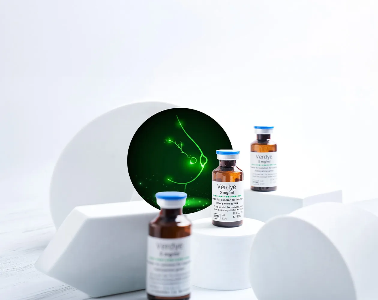
THE CURRENT STANDARD
A radiation-free, real-time fluorescence approach to intraoperative identification of sentinel lymph nodes and visualisation of lymphatic pathways in breast cancer.
Verdye is approved for the breast SLN indication across Austria, Belgium, Croatia, Czechia, Denmark, Finland, Germany, Greece, Hungary, Ireland, Italy, the Netherlands, Poland, Portugal, Romania, Slovenia, Spain, Sweden and the UK.
The presence of lymphatic metastases is an important prognostic factor for the survival of breast cancer patients, and their identification has a direct bearing on further treatment.1 Sentinel lymph node biopsy (SLNB) remains a cornerstone in the management of early breast cancer and is the current standard of care for clinically and radiologically node-negative patients. 2 Compared with axillary lymph node dissection, SLNB enhances a patient’s quality of life without compromising diagnostic accuracy or prognostic information.3
The traditional gold standard – radio-guided surgery with technetium-99m (Tc-99m) – reports SLN detection rates from 85.0% to 100%.4 Some clinicians add a blue dye to improve detection to 96.0%–99.8%,4,5 but blue dyes carry a recognised risk of skin and soft tissue necrosis, permanent injection-site tattooing and systemic reactions that limit routine use. 6,7,8,9,10 Tc-99m places an additional logistical burden on patients and hospitals: its use depends on access to nuclear medicine infrastructure, while pre-operative administration introduces additional appointments, departmental coordination and radiopharmaceutical handling requirements.
THE EVIDENCE FOR ICG
Clinical studies show that ICG fluorescence imaging achieves SLN detection rates comparable to established radioisotope and dual tracer techniques, 5,11,12,13 with some meta-analyses reporting higher detection rates than individual conventional tracers – including in patients who have received neoadjuvant chemotherapy. 4,12 ICG also maintains effectiveness in patients with a high body mass index, up to 40. 4,12,14
mean SLN detection rate across 2,168 patients 14
mean number of sentinel nodes detected per patient 14
transcutaneous imaging depth of lymphatic vessels with NIR camera systems15
single adverse reaction reported in Diagnostic Green pharmacovigilance data (25 Feb 2025)**

Meta-analysis of 2,168 patients undergoing ICG imaging procedures (5–10 mg per injection).14

REAL-WORLD EVIDENCE
A 2026 prospective single-centre UK cohort of 300 consecutive SLNB procedures across 292 patients – one of the largest evaluations of ICG as a sole tracer to date – reported a 99.7% nodal procedural detection rate (299/300 procedures), a 97.7% ICG fluorescent nodal detection rate (293/300 procedures), zero ICG-related adverse events or allergic reactions and a median operative time of 6.5 minutes from incision to first node identification. Detection was not significantly influenced by age, BMI, tumour size, or procedure type on multivariable analysis. 19
Procedures were performed by five different breast surgeons with varying levels of prior fluorescence-guided SLNB experience – including newly appointed consultants. Detection rates remained consistently high throughout, supporting the reproducibility of technique in routine clinical practice. 19

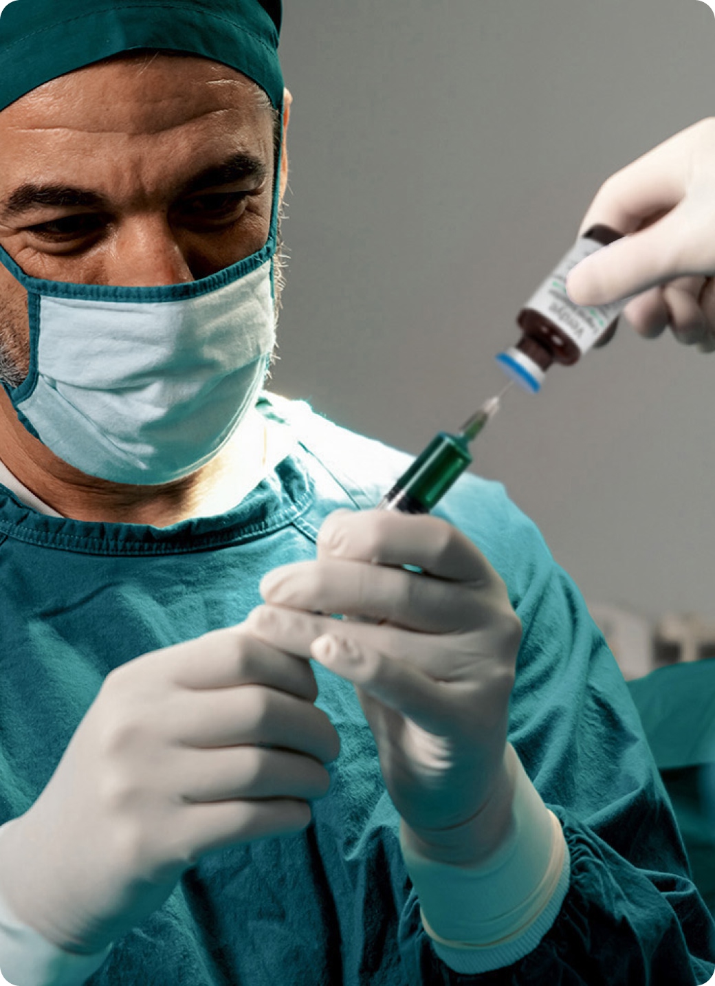
PROCEDURE
 Reconstitute Immediately prior to surgery, a 25 mg vial of ICG is prepared with 5 mL of water for injections.
InjectionOnce the patient is anaesthetised, 1–2 mL of ICG is injected intradermally, subcutaneously, or peritumourally into a region upstream of the lymph nodes of interest which drain that area.
Track in real timeHolding a near-infrared (NIR) camera system above the injected tissue, lymphatic vessels containing ICG are visible transcutaneously (to a depth of 1.0–1.5 cm 15) and can be mapped to the axilla, allowing real-time visualisation of lymph flow through the vessels.
Identify the nodeAfter the axilla is incised and dissected to the axillary fascia, the NIR camera is used to examine tissue fluorescence and identify the sentinel lymph node directly.
Reconstitute Immediately prior to surgery, a 25 mg vial of ICG is prepared with 5 mL of water for injections.
InjectionOnce the patient is anaesthetised, 1–2 mL of ICG is injected intradermally, subcutaneously, or peritumourally into a region upstream of the lymph nodes of interest which drain that area.
Track in real timeHolding a near-infrared (NIR) camera system above the injected tissue, lymphatic vessels containing ICG are visible transcutaneously (to a depth of 1.0–1.5 cm 15) and can be mapped to the axilla, allowing real-time visualisation of lymph flow through the vessels.
Identify the nodeAfter the axilla is incised and dissected to the axillary fascia, the NIR camera is used to examine tissue fluorescence and identify the sentinel lymph node directly.
GET STARTED
Unlike Tc-99m, ICG is prepared and injected after the patient is already anaesthetised in theatre – there is no separate nuclear medicine appointment to schedule the day before surgery.
WHY SURGEONS ARE SWITCHING
Provides real-time visualisation, allowing surgeons to precisely identify and remove SLNs during surgery.
Studies show ICG is superior or at least non-inferior to other tracers in identifying lymph nodes. 4,5,11,12,13 A small injection administered under anaesthesia minimises patient discomfort.
A more favourable safety profile compared with blue dyes, with only a single adverse reaction identified in 12,500 patients in pharmacovigilance data.**
Little or no tattooing with ICG, typically resolving within weeks – versus blue dye staining that can persist months to years, 16 and superparamagnetic iron oxide (SPIO) tracers reporting up to 37.8% immediate and 9.4% long-term (36-month) skin staining. 17
ICG does not cause MRI susceptibility artefacts. By contrast, superparamagnetic iron oxide (SPIO) tracers may produce persistent artefacts around the injection site and draining lymph nodes, potentially reducing image quality or making follow-up breast MRI difficult to interpret. 18
ICG is administered intraoperatively with a short interval before imaging and typically no need for massage of breast tissue. By comparison, SPIO labelling specifies a waiting period of at least 20 minutes and five minutes of vigorous massage before sentinel lymph node biopsy.18 Unlike Tc-99m, ICG does not require a separate pre-operative procedure or access to a nuclear medicine department.
POOLED CLINICAL EVIDENCE
Across nine independent meta-analyses, ICG shows a higher per-case SLN detection rate and higher sensitivity for cancer-positive nodes than Tc-99m, blue dye (BD), or the RI+BD combination – alongside the lowest false negative rate.
| COMPILED META-ANALYSIS RESULTS |
ICG | TC-99M | BLUE DYE | RI+BD |
|---|---|---|---|---|
| Allergic reactions | Very rare (<1/10,000) |
Erythema, urticaria, angioedema |
Soft tissue necrosis, metabolic & haematologic effects |
Allergic reactions related to both methods |
| Dosing timing | During surgery | Day before | During surgery | Requires administration before and during surgery |
| SLN detection rate | 97.8%* | 94.9% | 85.5% | 97.7% |
| Radiation exposure | None* | Yes | None | Yes |
| Pre-operative procedure | Not required* | Yes. Nuclear medicine infrastructure required. | Not required | Yes. Nuclear medicine infrastructure required. |
| Cancer(+ve) node sensitivity |
95.7%* | 91.2% | 83.1% | 94.2% |
| False negative rate | 4.3%* | 8.8% | 16.9% | 5.8% |
*Diagnostic Green, data on file
**Based on Diagnostic Green Pharmacovigilance data, 25 Feb 2025: a single adverse reaction was identified in 12,500 patients.
1. Goyal A, Newcombe RG, Chhabra A et al. Factors affecting failed localisation and false negative rates of sentinel node biopsy in breast cancer – results of the ALMANAC validation phase. Breast Cancer Res Treat 2006; 99: 203–208.
2. Rao R, Euhus D, Mayo HG et al. Axillary node interventions in breast cancer: a systematic review. JAMA 2013; 310: 1385–1394.
3. Cykowska A, Marano L, D’Ignazio A et al. New technologies in breast cancer sentinel lymph node biopsy; from the current gold standard to artificial intelligence. Surg Oncol. 2020 Sep;34:324–335.
4. Bargon CA, Huibers A, Young-Afat DA et al. Sentinel lymph node mapping in breast cancer patients through fluorescence imaging using Indocyanine green – the Influence Trial. Ann Surg. 2022 Nov 1;276(5):913–920.
5. Pitsinis V, Kanitkar R, Vinci A et al. Results of a Prospective Randomized Multicenter Study Comparing Indocyanine Green (ICG) Fluorescence Combined with a Standard Tracer Versus ICG Alone for Sentinel Lymph Node Biopsy in Early Breast Cancer: The INFLUENCE Trial. Ann Surg Oncol. 2024 Dec;31(13):8848–8855.
6. Mazouni C, Koual M, De Leeuw F et al. Prospective evaluation of limitations of near infrared imaging in detecting axillary sentinel lymph nodes in primary breast cancer. Breast J 2018; 24:1006–1009.
7. van der Horst JC, de Bock MJ, Klinkenbijl JH. Anaphylactic shock after injection of patent blue for sentinel lymph node biopsy. Ned Tijdschr Geneeskd. 2001 Oct 27;145(43):2086–8.
8. Hounschell C, Kilgore L, Pruitt P et al. Evaluation of learning curve with Indocyanine Green (ICG) versus blue dye for sentinel lymph node biopsy in breast cancer. Am J Surg. 2024 Jan;227:218–223.
9. Wilke LG, McCall LM, Posther KE et al. Surgical complications associated with sentinel lymph node biopsy: results from a prospective international cooperative group trial. Ann Surg Oncol. 2006 Apr;13(4):491–500.
10. Raut CP, Hunt KK, Akins JS et al. Incidence of anaphylactoid reactions to isosulfan blue dye during breast carcinoma lymphatic mapping in patients treated with preoperative prophylaxis. Cancer. 2005 Aug 15;104(4):692–9.
11. Kedrzycki MS, Leiloglou M, Ashrafian H et al. Meta-analysis Comparing Fluorescence Imaging with Radioisotope and Blue Dye-Guided Sentinel Node Identification for Breast Cancer Surgery. Ann Surg Oncol. 2021 Jul;28(7):3738–3748.
12. Staubach P, Scharl A, Ignatov A et al. Sentinel lymph node detection by means of indocyanine green using the Karl Storz VITOM® fluorescence camera: a comparison between primary sentinel lymph node biopsy versus sentinel lymph node biopsy after neoadjuvant chemotherapy. J Cancer Res Clin Oncol. 2021 Jun;147(6):1813–1823.
13. White KP, Sinagra D, Dip F et al. Indocyanine green fluorescence versus blue dye, technetium- 99M, and the dual-marker combination of technetium-99M + blue dye for sentinel lymph node detection in early breast cancer – meta-analysis including consistency analysis. Surgery. 2024 Apr;175(4):963–973.
14. Summary of Product Characteristics (SmPC). Indocyanine Green 5 mg/mL powder for solution for injection. Approved in the context of a European Mutual Recognition Procedure. Status: 06/2025.
15. Cornelissen AJM, van Mulken TJM, Graupner C et al. Near-infrared fluorescence image-guidance in plastic surgery: a systematic review. Eur J Plast Surg. 2018;41(3):269–278.
16. Gumus M, Gumus H, Jones SE et al. How long will I be blue? Prolonged skin staining following sentinel lymph node biopsy using intradermal patent blue dye. Breast Care (Basel). 2013 Jun;8(3):199–202.
17. FDA Summary of Safety and Effectiveness Data (SSED) for Magtrace®.
18. Per FDA-published Magtrace® labelling (IFU), FDA website (accessed 21 Aug 2025).
19. Pitsinis V, Khitab N, Jordan L, Vinci A, Kanitkar R, Elseedawy E, Smith J. ICG-only Sentinel Lymph Node Biopsy in Breast Cancer: A Prospective UK Cohort Demonstrating Consistent Detection Across Patient and Tumour Characteristics – A Follow-on to the INFLUENCE Trial. Ann Surg Oncol. 2026. doi:10.1245/s10434-026-20316-w.
GET STARTED