

CLINICAL APPLICATION Objective assessment in colorectal surgery
In colorectal and gastrointestinal surgery, adequate tissue perfusion is a critical consideration when constructing an anastomosis.
ICG fluorescence imaging enables surgeons to visualise perfusion in real time; however, interpretation is still largely visual and may vary depending on imaging distance, camera system, ICG timing, tissue characteristics and user experience.
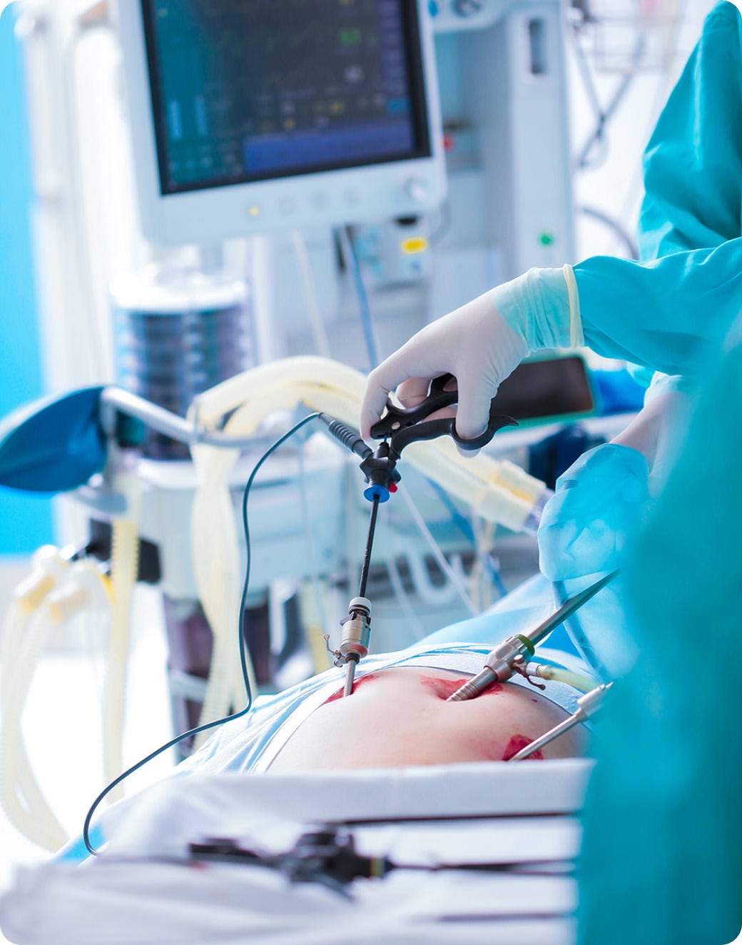
Note: PerfusionWorks does not replace surgical judgement. It is designed to complement surgeon expertise by adding objective quantitative information to the intraoperative assessment of tissue perfusion.
How it works
Clinical research increasingly highlights the potential value of moving beyond visual assessment alone towards quantitative fluorescence analysis. By converting fluorescence intensity over time into measurable perfusion parameters, quantification can support a more objective and reproducible approach to intraoperative perfusion assessment.
PerfusionWorks adds quantitative analysis to fluorescence-guided surgery by generating objective perfusion data from user-selected regions of interest. This enables surgeons to assess perfusion in a more structured way, alongside their clinical judgement and established surgical decision-making.
In colorectal surgery, PerfusionWorks may support:
-
 Objective assessment of perfusion at the planned transection or anastomotic site
-
Comparison of perfusion between selected tissue regions
-
Measurement-led assessment of fluorescence dynamics
-
More consistent intraoperative documentation
-
A structured approach to fluorescence-guided perfusion assessment
GET STARTED