

WHAT IT’S FOR Intended Use
The IC-Flow™ Imaging System is used to capture and view fluorescent images for various indications such as: visual assessment of blood flow, visual assessment of the lymphatic system, organ transplant procedures, plastic, micro and reconstructive surgery, visual assessment of the hepatobiliary system (liver tumours and liver metastases), and visual assessment of bile ducts during hepatobiliary surgery.
WHERE IT’S USED
The IC-Flow™ Imaging System is used as an adjunctive method for visualisation of ICG fluorescent images.
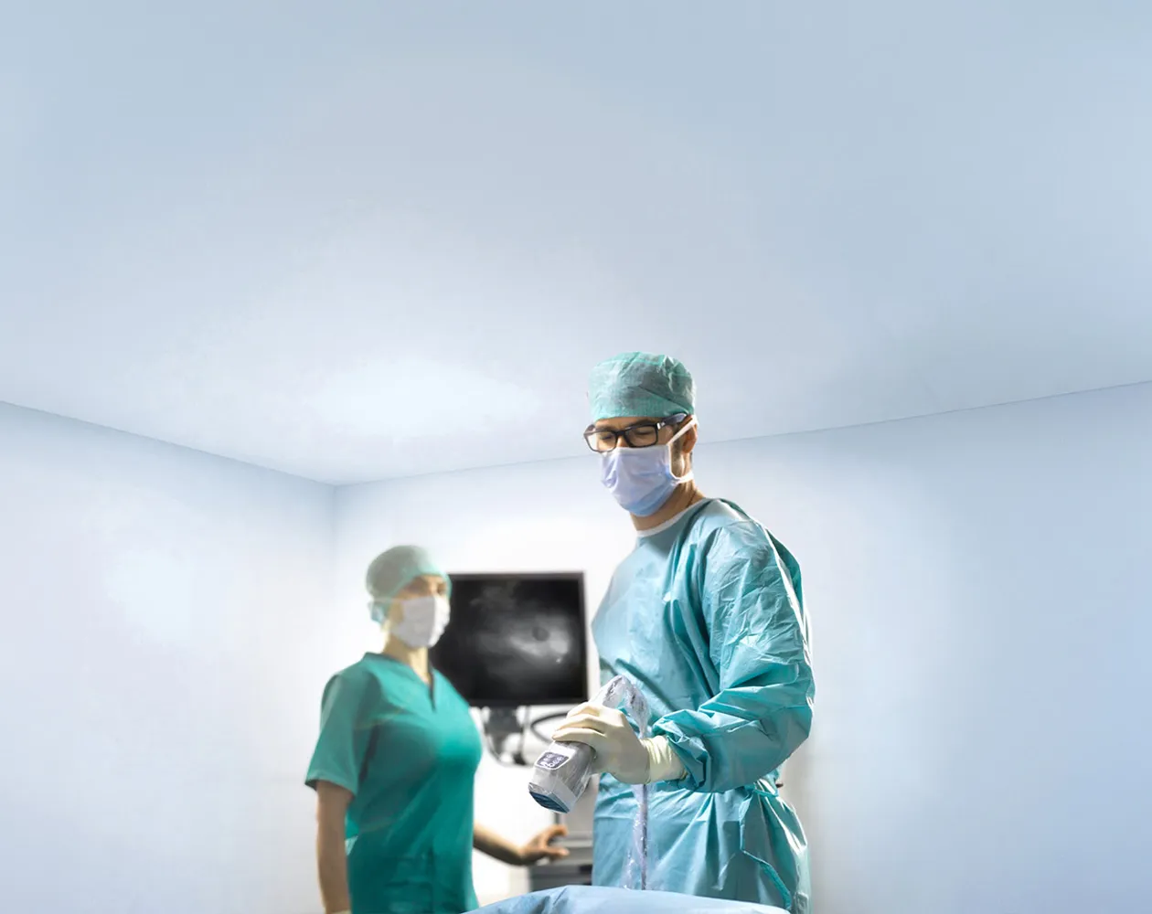
The IC-Flow™ Imaging System is intended for near-infrared fluorescence imaging in adult and paediatric patients one month of age and older. The IC-Flow™ Imaging System is intended for use by qualified surgeons, physicians or healthcare professionals in doctors’ offices and hospitals.
IC-Flow visualises blood flow as an adjunctive method for evaluating tissue perfusion. By capturing the ICG fluorescence signal in real time, surgeons can assess whether tissue is receiving adequate blood supply at key points during a procedure – and repeat that assessment as many times as needed, given ICG’s short half-life and IC-Flow’s fast, radiation-free imaging.
Perfusion assessment based on clinical judgement alone may miss early signs of compromised blood supply. Real-time fluorescence visualisation gives surgeons an additional, objective data point at the moment a decision needs to be made.
IC-Flow visualises lymphatic flow, supporting physicians in mapping and assessing the lymphatic system simply and effectively – in theatre or in an outpatient clinic setting.
Clear visualisation of lymphatic flow supports procedures such as lymphatic mapping, where identifying the pattern and integrity of lymphatic drainage is central to the clinical decision at hand.
IC-Flow visualises tissue-transfer circulation, supporting the intraoperative assessment of perfusion in organ transplant procedures.
Confirming adequate perfusion of transplanted tissue at the time of surgery – and being able to reassess quickly if a concern arises – provides additional visual information to inform intraoperative assessment before closure.
IC-Flow visualises tissue-transfer circulation in tissue and free flaps used in plastic, micro and reconstructive surgery, giving surgeons a real-time view of flap perfusion during the procedure.
Flap failure and necrosis carry significant clinical and financial cost. Published studies have reported lower complication rates with intraoperative ICG-based assessment than with clinical assessment alone in selected settings (see the Benefits page for cited outcomes in breast reconstruction and flap necrosis).
IC-Flow is CE-marked for the visual assessment of the hepatobiliary system, giving surgeons a real-time fluorescence view to support orientation during hepatobiliary procedures.
Anatomical variation in the biliary tree and liver vasculature is common. A real-time fluorescence view can give the surgical team an additional visual reference during dissection and reconstruction.
IC-Flow is CE-marked for the visual assessment of the hepatobiliary system, specifically including liver tumours and liver metastases.
Fluorescence-based visualisation has been used as an adjunct in hepatic surgery to support the intraoperative localisation of lesions, including small or subcapsular lesions that may be difficult to identify by palpation or inspection alone.
IC-Flow is CE-marked for the visual assessment of bile ducts during hepatobiliary surgery, giving surgeons real-time fluorescence visualisation to assist in identifying biliary anatomy during open hepatobiliary procedures.
Clear intraoperative visualisation of the biliary tree provides an additional anatomical reference during hepatobiliary surgery.
GET STARTED