

Fluorescence in Surgery Extending the surgical view
Fluorescence-guided surgery is changing what can be seen – and increasingly, what can be measured – during a procedure.

what is icg
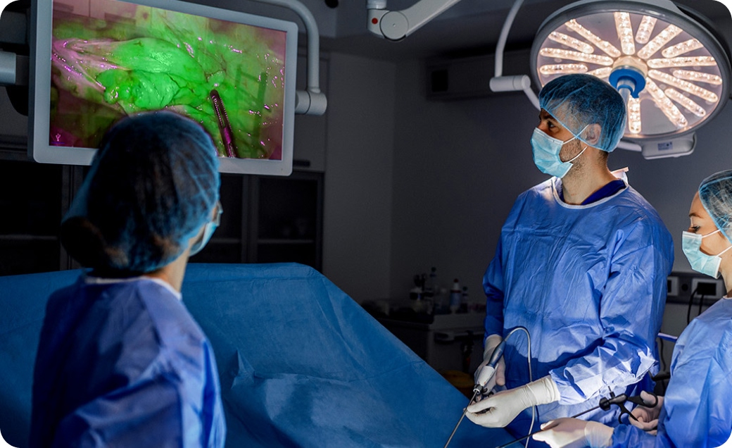
Using Indocyanine Green (ICG) with a compatible near-infrared imaging system can provide real-time visual information on selected anatomy and physiology alongside the conventional surgical view. It can allow surgeons to observe blood flow and tissue perfusion, trace lymphatic pathways and visualise relevant anatomical structures at critical points in a procedure.
First approved for clinical use in the United States in 1959, ICG has an established history in cardiac, circulatory, hepatic and ophthalmic diagnostics.1 Its evolution into surgery has been enabled by advances in near-infrared imaging, from standalone systems used in open procedures to integrated laparoscopic and robotic platforms.
Today, ICG fluorescence imaging sits at the convergence of pharmaceutical science, advanced imaging and digital surgery.
THE EVIDENCE
A growing clinical evidence base
65+
years of clinical use
20,000+
publications relating to ICG since 2000
3 Imaging environments
Open, laparoscopic and robotic imaging environments
The clinical literature around ICG continues to expand across gastrointestinal, hepatobiliary, vascular, oncological, plastic, reconstructive and other surgical fields. This growth reflects more than the versatility of the fluorescence agent. It also reflects a change in surgical imaging itself: from observing anatomy under white light to accessing real-time information on blood flow, tissue perfusion and lymphatic drainage within the operative field. The contribution of fluorescence nevertheless differs by application. It is most meaningful where it answers a defined clinical question – not simply where a fluorescence image can be produced.
Fluorescence angiography
Seeing blood flow differently
Assessment of tissue perfusion has traditionally relied on clinical indicators such as colour, pulsation, bleeding characteristics and the surgeon’s experience. ICG fluorescence angiography provides an additional dynamic view of the arrival and distribution of blood flow within tissue.
In colorectal surgery, a 2025 systematic review and meta-analysis included 45 studies and 14,333 patients. Overall anastomotic leak rates were 4.5% with ICG fluorescence angiography compared with 8.5% without it. The fluorescence assessment prompted a change in the surgical plan in 8.4% of cases in studies reporting this outcome.2
A separate 2025 health technology assessment concluded that adding ICG fluorescence imaging to visual assessment could reduce anastomotic leaks and reoperations, although the certainty of evidence varied between outcomes. Its model estimated that ICG fluorescence imaging could prevent 22 major anastomotic leaks per 1,000 patients undergoing colorectal surgery with an anastomosis.3
These findings illustrate an important distinction: fluorescence does not make the surgical decision. It provides additional information that may confirm the surgeon’s assessment—or identify a perfusion pattern that warrants reconsideration.

White light

ICG injection

Near infrared

Perfusion map

Clinical interpretation
In simple terms, these results correspond to approximately 42% lower odds of mastectomy skin-flap necrosis, 69% lower odds of fat necrosis and 60% lower odds of reoperation compared with conventional clinical assessment alone. Note these are relative reduction in odds not absolute reductions in complication rates.4 Seven studies also reported cost savings in flap surgery and breast reconstruction.4
THE EVIDENCE
What is Fluorescence-Guided Surgery?
In plastic and reconstructive surgery, the viability of a flap depends on adequate tissue perfusion. ICG fluorescence angiography can help identify areas of otherwise unsuspected hypoperfusion before the procedure is completed.
A 2024 systematic review and meta-analysis included 25 studies, with ICG fluorescence angiography used predominantly in breast surgery and head and neck reconstruction. In breast procedures, its use was associated with lower odds of:
-
 Mastectomy skin-flap necrosis: OR 0.58
-
Fat necrosis: OR 0.31
-
Reoperation: OR 0.40

LYMPHATIC IMAGING
Mapping lymphatic pathways
Following local administration, where approved, ICG fluorescence can enable lymphatic drainage pathways and sentinel lymph nodes to be visualised in real time. In breast cancer surgery, a network meta-analysis of 35 studies found that ICG achieved better sentinel lymph node detection than blue dye alone and performance comparable to established dual-tracer techniques.5
Fluorescence also allows the surgeon to see the lymphatic pathway directly during the procedure. Where supported by approved indications and local protocols, an ICG-based pathway may reduce dependence on radioactive tracers and associated nuclear-medicine scheduling. The clinical, operational and economic implications will vary between healthcare systems and local protocols.
Part of a multimodal pathway
Designed for the way surgery is performed
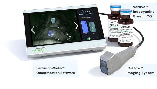
Fluorescence imaging has developed alongside open, laparoscopic and robotic surgery. In open procedures, systems such as IC-Flow™ can bring near-infrared fluorescence directly to the operative field. In laparoscopic surgery, fluorescence has become integrated into the minimally invasive camera view. Robotic platforms take this further by incorporating fluorescence within an increasingly digital surgical environment.
The imaging platform matters – a 2024 technical review identified significant variation between fluorescence systems in sensitivity, fluorescence intensity, image presentation and technical performance.6 What appears on screen is shaped not only by ICG, but also by the product and dose used, administration route and timing, patient physiology, tissue characteristics, imaging distance, ambient conditions and system performance.
There is therefore no universal indocyanine green dose for surgery. The appropriate ICG dose – including for colorectal, robotic or other procedures – depends on the approved product information, intended clinical application, route of administration and compatible imaging system.
THE NEXT STEP
From visual fluorescence to quantification
Most intraoperative fluorescence is still interpreted visually. The surgeon observes where fluorescence appears, how quickly it develops and how it is distributed across the tissue. However, fluorescence videos contains more information than a single image can convey. Its temporal and spatial characteristics can potentially be analysed to measure:
- Fluorescence arrival and inflow
- Time to maximum fluorescence
- Relative fluorescence intensity
- Wash-in and wash-out behaviour
- Variation between different regions of tissue
Published reviews have identified numerous approaches to fluorescence quantification, but also substantial differences in acquisition protocols, terminology, analytical methods and proposed thresholds.7,8 Progress therefore depends on more than producing a numerical value. Measurements must be reproducible, clinically validated and robust to camera, tissue and instrument movement.


LOOKING AHEAD
Fluorescence, robotics and AI
As fluorescence becomes more closely integrated with laparoscopic and robotic platforms, surgical video becomes a potential source of structured physiological data. Artificial intelligence and machine learning are being investigated for tissue recognition, image registration, motion correction, fluorescence-curve analysis and identification of spatial perfusion differences. Early research has demonstrated the feasibility of combining fluorescence analysis, biophysical modelling and machine
learning to assess anastomotic blood supply.9
These technologies remain in development and require rigorous clinical and regulatory validation. They nevertheless indicate the direction of travel: from a fluorescence image interpreted at a single moment towards reproducible measurements and clinically meaningful, data-driven decision support.
PERFUSIONWORKS™
Beyond visual fluorescence
PerfusionWorks™ has been developed to advance fluorescence imaging through quantification and data-driven decision support.
TRUST. INSIGHT. PRECISION.
From seeing fluorescence to understanding the signal
Fluorescence-guided surgery has progressed from standalone imaging to integration within open, laparoscopic and robotic procedures.
Across this evolution, its purpose has remained consistent: to provide the surgical team with additional visual information when it matters.
The next stage is not simply to generate more images. It is to make fluorescence information more consistent, measurable and clinically meaningful.
Diagnostic Green, through Verdye®, IC-GREEN®, IC-Flow™ and PerfusionWorks™, is working across this continuum – from the established fluorescence agent to imaging, quantification and the future of data-supported surgery.
REFERENCES
- US Food and Drug Administration. IC-GREEN® (indocyanine green for injection) prescribing information. Initial US approval: 1959. Revised 2024.
- McEntee PD, Singaravelu A, McCarrick CA, et al. Quantification of indocyanine green fluorescence angiography in colorectal surgery: a systematic review of the literature. Surg Endosc. 2025;39(4):2677–2691. doi:10.1007/s00464-025-11621-8.
- Ontario Health (Quality). Indocyanine green fluorescence imaging for colorectal surgery: a health technology assessment. Ont Health Technol Assess Ser. 2025;25(3):1–124. PMID: 40727261.
- Singaravelu A, McCarrick C, Potter S, Cahill RA. Clinical and cost-effectiveness of intraoperative flap perfusion assessment with indocyanine green fluorescence angiography in breast and head and neck reconstructions: a systematic review and meta-analysis. Microsurgery. 2024;44(8):e31250. doi:10.1002/micr.31250.
- Mok CW, Tan SM, Zheng Q, Shi L. Network meta-analysis of novel and conventional sentinel lymph node biopsy techniques in breast cancer. BJS Open. 2019;3(4):445–452. doi:10.1002/bjs5.50157.
- Preziosi A, Cirelli C, Waterhouse D, et al. State of the art medical devices for fluorescence-guided surgery: technical review and future developments. Surg Endosc. 2024;38(11):6227–6236. doi:10.1007/s00464-024-11236-5.
- Slooter MD, Eshuis WJ, Cuesta MA, et al. Defining indocyanine green fluorescence to assess anastomotic perfusion during gastrointestinal surgery: a systematic review. Br J Surg. 2021;108(11):1301–1310.
- McEntee PD, et al. Quantification of indocyanine green fluorescence angiography in colorectal surgery: a systematic review. Surg Endosc. 2025.
- He W, Zhu H, Rao X, et al. Biophysical modeling and artificial intelligence for quantitative assessment of anastomotic blood supply in laparoscopic low anterior rectal resection. Surg Endosc. 2025;39(5):3412–3421. doi:10.1007/s00464-025-11693-6.
Publication count based on a PubMed search for ICG-related publications from January 2000 to July 2026. Approved indications, administration routes and dosing vary by product and territory. Healthcare professionals should consult the locally approved product information before use.