

VERDYE APPLICATIONS Verdye™ (Indocyanine Green, ICG) in Liver Function Diagnostics
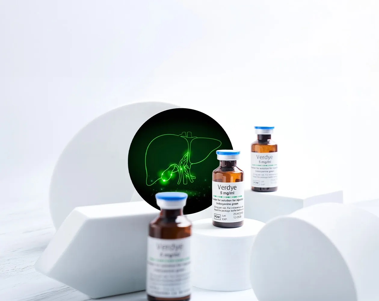
Approved INDICATIONS
A dynamic measure of how the liver is functioning
Blood tests provide important information about liver injury and biochemical status. Verdye™ (indocyanine green, ICG) enables something different: a dynamic, quantitative assessment of the liver’s ability to receive, extract and excrete ICG.
Verdye is indicated for:
Measurement of the excretory function of the liver¹

MEASUREMENT PRINCIPLE
Measurement of liver blood flow
Following intravenous administration, ICG binds extensively to plasma proteins and remains predominantly within the vascular compartment. Its concentration and rate of disappearance from the circulation can be measured using an appropriate validated method to support the quantitative assessment of liver blood flow.¹˒²
MEASUREMENT PRINCIPLE
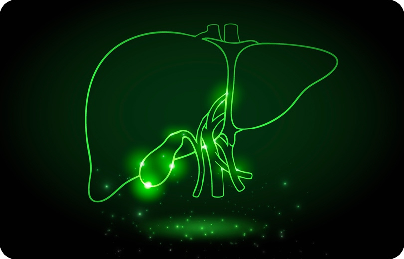
Measurement of the excretory function of the liver
ICG is taken up from the blood by the liver and excreted into the bile. It does not undergo significant extrahepatic or enterohepatic circulation.¹˒² Measuring how rapidly ICG disappears from the bloodstream therefore provides a dynamic assessment of hepatic uptake and excretory function.
MEASURED PARAMETERS
From administration to quantitative result
Following intravenous administration, the change in circulating ICG concentration can be measured over time. Depending on the selected method, the resulting parameters may include:¹
ICG PLASMA DISAPPEARANCE RATE (ICG-PDR):
the rate at which ICG is removed from the bloodstream
PLASMA HALF-LIFE:
the time required for the circulating concentration of ICG to decrease by half
ICG RETENTION AT 15 MINUTES (ICG-R15):
the proportion of ICG remaining in the circulation 15 minutes after administration
CLEARANCE:
the volume of plasma from which ICG is removed over a defined period
Together, these measurements provide quantitative information about liver function at the time the test is performed.

STATIC VS DYNAMIC
Dynamic assessment beyond static biochemical markers
Routine biochemical tests measure the concentration of individual markers at a particular point in time. ICG clearance measures a physiological process: the uptake and excretion of ICG by the liver.²
This makes it a valuable complementary measure when a dynamic assessment of hepatic function is required.³
Static Assessment
- Blood Tests
- Measures biochemical markers
- Single point in time
VS
Dynamic Assessment
- Verdye
- Measures physiological function
- Real-time hepatic uptake and excretion
THE EVIDENCE BASE
Supported by extensive clinical investigation
The use of ICG clearance as a quantitative liver-function test is supported by decades of clinical research.
698 patients:
ICG clearance associated with liver dysfunction
In a study of 698 patients undergoing liver resection, both preoperative ICG-PDR and ICG-R15 were significantly associated with postoperative liver dysfunction. Patients with impaired ICG clearance were approximately twice as likely to develop liver dysfunction and experienced more severe complications and longer hospital stays.⁴
4,852 patients:
evidence across 17 studies
A 2022 systematic review and meta-analysis examined 17 studies involving 4,852 patients.
The review confirmed that ICG clearance is one of the most widely used dynamic assessments in the preoperative evaluation of liver function.
It also concluded that ICG clearance should form part of a broader, multiparametric assessment rather than being used as a standalone predictor of postoperative liver failure.³
MEASUREMENT METHODS
Flexible measurement approaches
Depending on the equipment and methodology used, ICG kinetics may be measured:¹
-
 Non-invasivelyusing pulse dye densitometry or near-infrared spectroscopy
-
 Invasivelyusing suitable fibre-optic probes or intravascular catheters
-
 ConventionallyBy determining ICG concentration in serial blood samples
Parameters that can be obtained from the passage of Verdye through the circulation include:
- Area under the first-pass curve
- Transit time
- Half-life
- Plasma disappearance rate
- Retention rate¹
The administration site, measurement equipment and analytical method are important to the quality and interpretation of the results.

IMPORTANT INFORMATION
Always consult the locally approved Summary of Product Characteristics or prescribing information before use. Approved indications, presentations, dosing and instructions may vary by territory. ICG clearance results should be interpreted in conjunction with other relevant clinical information.
References
- Diagnostic Green Limited. Verdye 5 mg/ml powder for solution for injection: Summary of Product Characteristics. Revised June 2025. https://www.hpra.ie/find-a-medicine/for-human-use/authorised-medicines/details/38844
- Imai T, et al. Measurement of cardiac output by pulse dye densitometry using indocyanine green: a comparison with the thermodilution method. Anesthesiology. 1997;87(4):816–822. https://pubmed.ncbi.nlm.nih.gov/9357883/
- Iijima T, et al. Cardiac output and circulating blood volume analysis by pulse dye-densitometry. Journal of Clinical Monitoring. 1997;13(2):81–89. https://pubmed.ncbi.nlm.nih.gov/9112203/
- Hofer CK, et al. Pulsed dye densitometry with two different sensor types for cardiac output measurement after cardiac surgery: a comparison with the thermodilution technique. Acta Anaesthesiologica Scandinavica. 2004;48(5):653–657. https://pubmed.ncbi.nlm.nih.gov/15101865/
- Iijima T, et al. Circulating blood volume measured by pulse dye-densitometry: comparison with the conventional method using radioiodinated human serum albumin. Anesthesiology. 1998;89(6):1329–1335. https://pubmed.ncbi.nlm.nih.gov/9856706/
- Haruna M, et al. Blood volume measurement at the bedside using ICG pulse spectrophotometry. Anesthesiology. 1998;89(6):1322–1328. https://pubmed.ncbi.nlm.nih.gov/9856705/
- Imai T, et al. Accuracy and repeatability of blood volume measurement by pulse dye densitometry compared with the conventional method using chromium-51-labelled red blood cells. Intensive Care Medicine. 2000;26(9):1343–1349. https://pubmed.ncbi.nlm.nih.gov/11089762/
- Haller M, et al. Determination of plasma volume by indocyanine green in man: validation of the method and use in patients after cardiopulmonary bypass. British Journal of Anaesthesia. 1993;70(6):672–676. https://pubmed.ncbi.nlm.nih.gov/11430552/
- Roberts I, et al. Estimation of cerebral blood flow with near-infrared spectroscopy and indocyanine green. Lancet. 1993;342(8884):1425. https://pubmed.ncbi.nlm.nih.gov/7901707/
- Gora F, et al. Non-invasive measurement of cerebral blood flow in adults using near-infrared spectroscopy and indocyanine green: a pilot study. Journal of Neurosurgical Anesthesiology. 2002;14(3):218–222. https://pubmed.ncbi.nlm.nih.gov/12172295/
- Keller E, et al. Non-invasive measurement of regional cerebral blood flow and regional cerebral blood volume by near-infrared spectroscopy and indocyanine green dye dilution. NeuroImage. 2003;20(2):828–839. https://pubmed.ncbi.nlm.nih.gov/14568455/
- Milej D, et al. Quantification of cerebral blood flow in adults by dynamic contrast-enhanced near-infrared spectroscopy: validation against MRI. Journal of Cerebral Blood Flow & Metabolism. 2020;40(8):1672–1684. https://pubmed.ncbi.nlm.nih.gov/31500522/
GET STARTED