
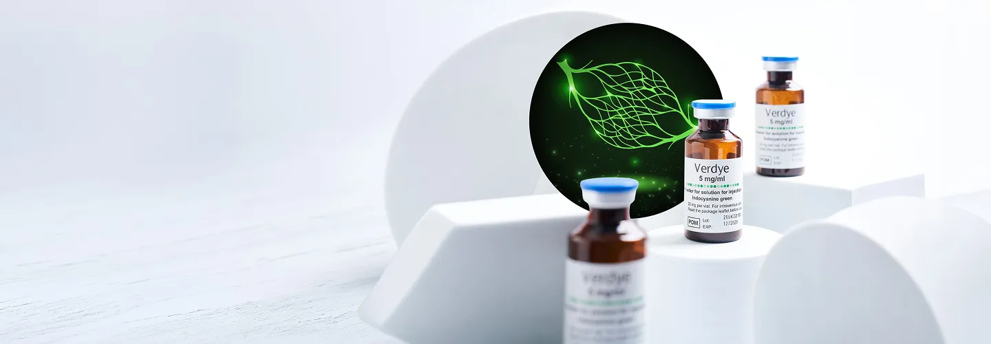
VERDYE™ APPLICATIONS Cardiac, circulatory and microcirculatory diagnostics
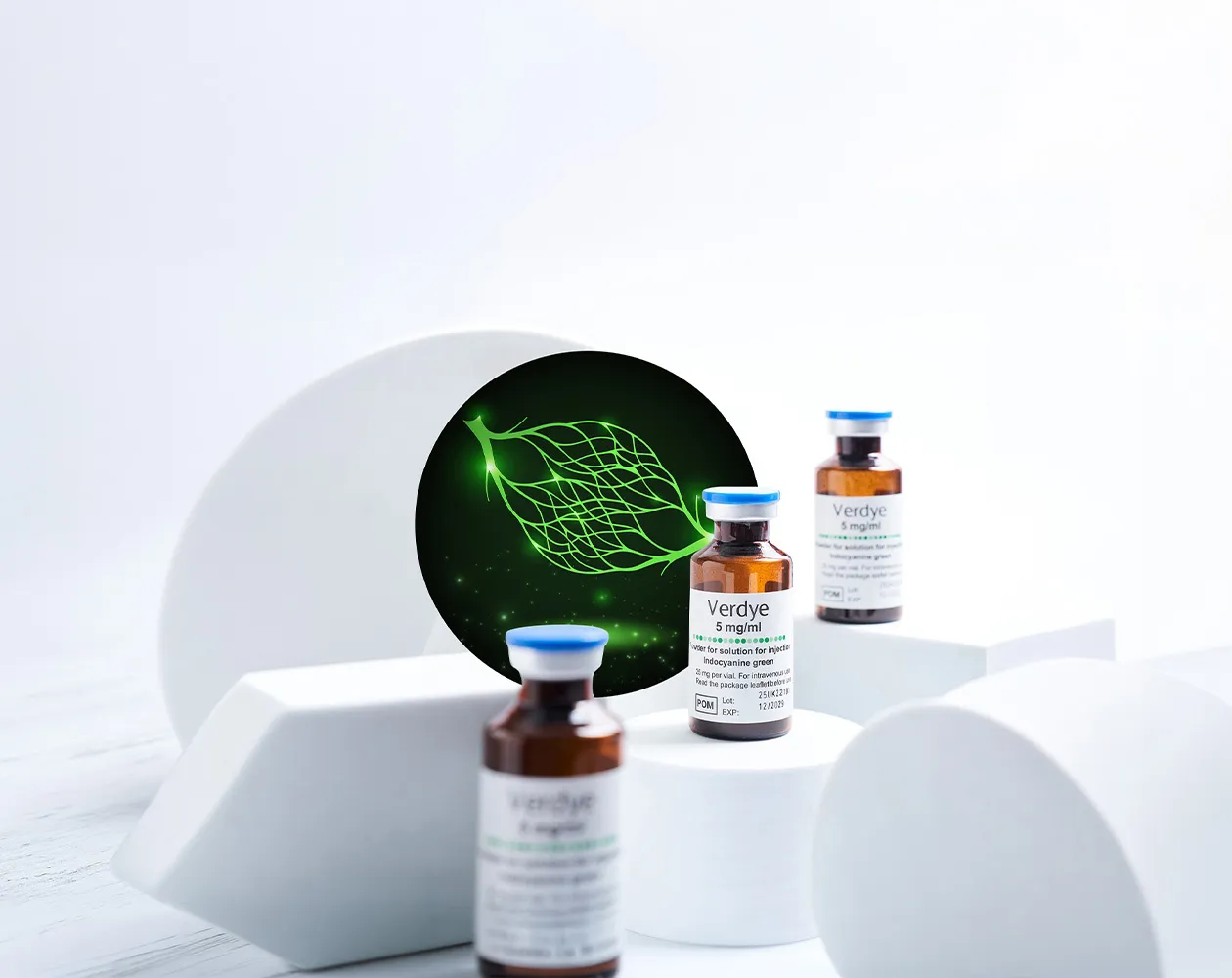
AUTHORISED INDICATIONS
Measuring cardiovascular function with Verdye
Cardiac output, stroke volume, circulating blood volume and cerebral perfusion provide important information about cardiovascular function and the movement of blood through the body.
VerdyeTM (Indocyanine Green, ICG) is authorised for:
Measurement of circulating blood volumes
Measurement of cerebral perfusion¹

Following intravenous administration, indocyanine green rapidly binds to plasma proteins and remains predominantly within the vascular compartment. This allows its passage through the circulation to be detected and analysed using indicator-dilution techniques.¹
CARDIAC PERFORMANCE
Measurement of cardiac output and stroke volume
Cardiac output is the volume of blood pumped by the heart each minute. Stroke volume is the amount of blood ejected with each contraction. Together, these measurements provide information about the heart’s pumping performance and the delivery of blood to the systemic circulation.
Following intravenous administration of Verdye, the first passage of indocyanine green through the circulation can be recorded as an indicator-dilution curve. The concentration of the indicator over time can then be used to calculate cardiac output using a compatible measurement and analysis system.¹
Supporting evidence
Clinical studies have evaluated ICG-based cardiac-output measurement against established methods:
In patients undergoing major surgery, Imai and colleagues reported that pulse dye densitometry using ICG provided repeated cardiac-output measurements with a degree of accuracy comparable to thermodilution.²
Iijima and colleagues evaluated ICG pulse dye densitometry for cardiac output and circulating blood volume. Cardiac-output results were compared with conventional dye-dilution and thermodilution measurements, including in patients undergoing coronary artery bypass surgery.³
The method has also been studied in patients following cardiac surgery, although the performance of individual measurement systems and sensor technologies has varied.⁴
The evidence supports ICG as an established indicator for cardiac-output measurement. Measurement performance depends on the equipment, sensor, administration technique and clinical conditions.

NEUROVASCULAR ASSESSMENT
Measurement of cerebral perfusion
Cerebral perfusion describes the delivery of blood to brain tissue. Its measurement provides information about cerebral blood flow and may be relevant where systemic or regional circulation is altered. Near-infrared spectroscopy can be used to detect changes in indocyanine green concentration as an intravenous bolus passes through the cerebral circulation. The resulting concentration-time curve can be analysed to estimate cerebral blood flow.¹
Supporting evidence
ICG has been investigated as an intravascular contrast indicator for cerebral perfusion measurement:
Roberts and colleagues demonstrated the use of near-infrared spectroscopy and intravenous ICG to estimate cerebral blood flow.⁹
Gora and colleagues reported the feasibility of non-invasive cerebral blood-flow measurement in adults following an intravenous bolus of ICG.10
Keller and colleagues used near-infrared spectroscopy with ICG dilution to measure regional cerebral blood flow and regional cerebral blood volume non-invasively.¹¹
In a later validation study, Milej and colleagues compared dynamic contrast-enhanced near-infrared spectroscopy using ICG with arterial-spin-labelling MRI. The measurements were strongly correlated, withWith an R2 of 0.88.¹²
This research supports the scientific basis for ICG-based cerebral perfusion measurement. The method requires compatible near-infrared detection and appropriate analysis technology.
MEASUREMENT METHODS
How Verdye is measured
The Verdye Summary of Product Characteristics describes several methods for cardiac, circulatory and cerebral blood-flow measurement:
-
 Non-invasivelyusing pulse dye densitometry or near-infrared spectroscopy
-
 Invasivelyusing fibre-optic probes or catheters in suitable vessels
-
 Conventionallythrough continuous blood withdrawal or collection of blood samples followed by photometric analysis¹
Parameters that can be obtained from the passage of Verdye through the circulation include:
- Area under the first-pass curve
- Transit time
- Half-life
- Plasma disappearance rate
- Retention rate¹
The administration site, measurement equipment and analytical method are important to the quality and interpretation of the results.
PRODUCT CHARACTERISTICS
Why Verdye is suited to circulatory measurement
Verdye has characteristics that support its use as an intravascular indicator:
- Approximately 95% binding to plasma proteins
- Volume of distribution corresponding to blood volume
- Defined near-infrared absorption peak at approximately 800nm
- Measurement at 800nm independently of changes in blood oxygen saturation
- Initial plasma elimination half-life of approximately 3–4 minutes
- Negligible renal, peripheral and pulmonary uptake¹
Indocyanine green is taken up almost exclusively from the plasma by the liver and excreted unchanged into the bile.¹

IMPORTANT INFORMATION
Verdye is a diagnostic medicinal product and should be administered under the supervision of a physician. The performance and clinical interpretation of any measurement depend on the compatible equipment, analytical method, administration technique and individual patient circumstances. Authorised indications and product information may differ between territories. Healthcare professionals should consult the applicable local Summary of Product Characteristics for complete information on dosing, administration, contraindications, warnings and precautions.
References
- Diagnostic Green Limited. Verdye 5 mg/ml powder for solution for injection: Summary of Product Characteristics. Revised June 2025. https://www.hpra.ie/find-a-medicine/for-human-use/authorised-medicines/details/38844
- Imai T, et al. Measurement of cardiac output by pulse dye densitometry using indocyanine green: a comparison with the thermodilution method. Anesthesiology. 1997;87(4):816–822. https://pubmed.ncbi.nlm.nih.gov/9357883/
- Iijima T, et al. Cardiac output and circulating blood volume analysis by pulse dye-densitometry. Journal of Clinical Monitoring. 1997;13(2):81–89. https://pubmed.ncbi.nlm.nih.gov/9112203/
- Hofer CK, et al. Pulsed dye densitometry with two different sensor types for cardiac output measurement after cardiac surgery: a comparison with the thermodilution technique. Acta Anaesthesiologica Scandinavica. 2004;48(5):653–657. https://pubmed.ncbi.nlm.nih.gov/15101865/
- Iijima T, et al. Circulating blood volume measured by pulse dye-densitometry: comparison with the conventional method using radioiodinated human serum albumin. Anesthesiology. 1998;89(6):1329–1335. https://pubmed.ncbi.nlm.nih.gov/9856706/
- Haruna M, et al. Blood volume measurement at the bedside using ICG pulse spectrophotometry. Anesthesiology. 1998;89(6):1322–1328. https://pubmed.ncbi.nlm.nih.gov/9856705/
- Imai T, et al. Accuracy and repeatability of blood volume measurement by pulse dye densitometry compared with the conventional method using chromium-51-labelled red blood cells. Intensive Care Medicine. 2000;26(9):1343–1349. https://pubmed.ncbi.nlm.nih.gov/11089762/
- Haller M, et al. Determination of plasma volume by indocyanine green in man: validation of the method and use in patients after cardiopulmonary bypass. British Journal of Anaesthesia. 1993;70(6):672–676. https://pubmed.ncbi.nlm.nih.gov/11430552/
- Roberts I, et al. Estimation of cerebral blood flow with near-infrared spectroscopy and indocyanine green. Lancet. 1993;342(8884):1425. https://pubmed.ncbi.nlm.nih.gov/7901707/
- Gora F, et al. Non-invasive measurement of cerebral blood flow in adults using near-infrared spectroscopy and indocyanine green: a pilot study. Journal of Neurosurgical Anesthesiology. 2002;14(3):218–222. https://pubmed.ncbi.nlm.nih.gov/12172295/
- Keller E, et al. Non-invasive measurement of regional cerebral blood flow and regional cerebral blood volume by near-infrared spectroscopy and indocyanine green dye dilution. NeuroImage. 2003;20(2):828–839. https://pubmed.ncbi.nlm.nih.gov/14568455/
- Milej D, et al. Quantification of cerebral blood flow in adults by dynamic contrast-enhanced near-infrared spectroscopy: validation against MRI. Journal of Cerebral Blood Flow & Metabolism. 2020;40(8):1672–1684. https://pubmed.ncbi.nlm.nih.gov/31500522/
GET STARTED